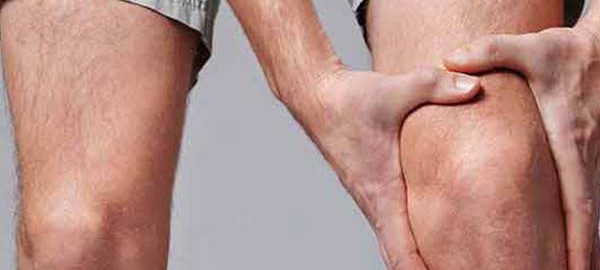
Tag Archives: orthopaedic in Dwarka

Cuboid

Leg Injuries – Cuboid Syndrome

Cuboid syndrome is the result of injuries to the joints and ligaments surrounding the cuboid bone. This is one of the seven tarsal bones of the foot (talus or astragalus, calcaneus, navicular, cuboid and 3 cuneiform bones).

From a medical point of view, the condition is called a cuboid subluxation and occurs when the cuboid bone moves down and out, without being aligned with the calcaneus bone.

The cuboid syndrome causes pain in the external side of the foot, which can be confused with other conditions.

Clinical picture

Cuboid syndrome encompasses a variety of common symptoms and other conditions, sometimes making it difficult to diagnose. The most common symptoms include:

  • Pain on the outside of the foot, which affects including fingers 4 and 5;
  • Pain that aggravates the standing state, due to body weight ;
  • Painful pain, dry or sharp and acute;
  • Weakness and difficulty in walking and jumping;
  • Redness, inflammation, and sensitivity in the injured area;
  • Walking painkillers, to reduce pain;
  • Reducing the range of movements of the foot and ankle.

Studies have reported that, in general, the cuboid syndrome is not uncommon in the population, but occurs most commonly in athletes and dancers. When the cuboid syndrome is correctly identified and treated, most people experience a complete recovery.

Causes

Cuboid syndrome can have different causes, but the most common are:

  • Overload – this condition, along with injury, is most commonly seen in athletes and dancers. These groups of athletes tend to have intense physical activity, regardless of the presence of pain. Overload injuries tend to develop after long periods of intense activity, such as running;
  • Ankle dislocation – ankle dislocation can also lead to injury to the Tarsus bones of the foot. A specialist study reported that about 40% of people with ankle dislocations or sprains can develop cuboid syndrome;
  • Foot walking in pronation – people who develop this type of walking have a higher risk of having the cuboid syndrome because the muscles of the leg can pull the tarsal bones into abnormal positions;
  • Other causes – climbing, uneven surfaces and sports involving fast and lateral movements, such as tennis, can cause the cuboid syndrome. Wearing shoes of poor quality or inadequate sizes may be another reason.

Also, if you do not take into account the indications of orthopaedic in Delhi regarding rest and recovery from a leg injury, various complications, including cuboid syndrome, may occur.

Diagnostics
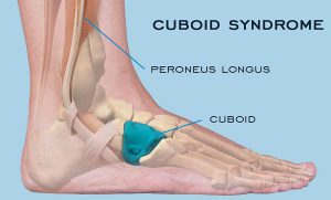

The foot represents a complex, flexible and resistant segment of the body. It contains about 100 muscles, ligaments and tendons, 28 bones and 30 joints.

The complex structure of the foot and the non-specific nature of the pain within the cuboid syndrome make this injury difficult to diagnose.

Sometimes, medical imaging techniques such as X-ray or magnetic resonance imaging (MRI) do not identify signs of this condition, even if it is present. The cuboid syndrome may also mimic the symptoms of other foot problems, such as fractures or sprained heels.

In order to establish a correct diagnosis and to find the most effective treatment, the physician must perform a thorough physical examination and examine a person’s medical history.

Treatment options

Treating cuboid syndrome always involves rest and elimination of activities that involve putting pressure on the injured leg. Applying ice and an elastic phase, but also raising the foot above the level of the heart on a pillow are other useful methods.

If the pain persists or worsens, the orthopaedic in Dwarka should be consulted urgently. Certain maneuvers performed by the therapists in the field can help to improve or eliminate the symptoms.

These maneuvers are meant to force the bone to return to its original position. However, it should be noted that such maneuvers are not recommended if there are other associated conditions, such as arthritis, bone disease, fractures, circulatory or nerve disorders.

Additional treatments may include the use of orthotics to support and properly align the foot, but also to administer anti-inflammatory drugs to reduce pain and inflammation.

In some cases, leg massage can help to reposition the bone. Surgery is used in rare cases when other treatment options have not worked.

Recovery

The length of time required for recovery from cuboid syndrome depends on several factors, such as:

  • Duration of the treatment until the start of treatment;
  • Cause of cuboid syndrome ;
  • If the initial injury was mild or severe;
  • If the instructions of orthopaedic in West Delhi were taken into account;
  • If the condition has been correctly diagnosed and treated.

Physiotherapy in Dwarka can play an important role in the complete recovery of a person with the cuboid syndrome. Also, this therapy can prevent further complications.

Physical therapy includes:

  • Reconsolidation of the strength and function of the foot;
  • Maintenance of the muscles;
  • Exercises to improve balance.

In some cases, to avoid pressure on the injured leg, the orthopaedic surgeon in Delhi may indicate the use of squats or walking sticks.

Risk factors

The common risk factors for the cuboid syndrome are:

  • Overweight or obesity;
  • Wearing shoes of inappropriate sizes;
  • Wearing pairs of shoes of inferior quality;
  • Practicing physical exercises without adequate heating;
  • Practicing physical activities without taking into account pain and without having sufficient rest periods;
  • Performing activities, such as running or walking on uneven or rough surfaces;
  • Fractures of neighboring bones of the cuboid;
  • Practicing sports such as athletics or ballet;
  • Practicing physical activities that involve repetitive movements and increased impact on the foot;
  • Association of other conditions such as arthritis or osteoporosis.

Perspective

Cuboid syndrome is not a serious condition and can be easily treated by the orthopaedic surgeon in Dwarka, sometimes involving physical therapy. The perspective is usually very good for people with this condition. Following treatment, most people can return to normal activities, with little risk of recurrence.

The orthopedic in Delhi should be notified if persistent pain occurs in the foot before doing any maneuver that could aggravate the situation.

Often there may be other health conditions that mimic cuboid syndrome. Rapid identification and early treatment of health status offer the best chances of recovery.

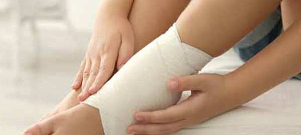
meniscus lesions of knee joint

Meniscus Lesions of The Knee Joint

The two meniscus can be easily injured by a rotation of the knee during a sustained effort. Partial or total meniscus rupture occurs at an abrupt torsion or thigh rotation, while the foot stays in place (for example, at a sudden turn to hit the tennis ball).

If the rupture is minimal, the meniscus remains connected in the anterior and posterior part of the knee; if the rupture is large, the meniscus may hang from a cartilage filament. The severity of a rupture depends on the location and extent.

Symptoms

In general, pain in the meniscus rupture occurs, especially when the leg is straightened (at the extension of the leg). The pain may be moderate and the patient may continue the activity.

Severe pain occurs when a fragment of the meniscus remains between the femur and tibia. Swelling may occur immediately after injury if the blood vessels are damaged, or a few hours after injury if the joint space is filled with fluid produced in response to inflammation. If the synovial envelope is damaged, it becomes inflamed and produces fluid to protect itself, installing the synovial envelope syndrome.

These changes cause swelling of the knee. Sometimes an untreated injury can become painful after several months or even years later, especially if the knee has been injured a second time. After an injury, the knee may become blocked or weakened. Symptoms of a meniscus lesion may resolve spontaneously, but frequently the symptoms persist and require treatment.

Diagnostic

In addition to the patient’s anamnesis, which describes the onset of pain and swelling, the orthopaedic in Dwarka may perform an x-ray of the affected knee. The examination may include a test in which the doctor flexes the leg, then rotates internally and externally while extending the leg. The pain from this maneuver suggests the meniscus rupture. An imaging magnetic resonance test may be recommended to confirm the diagnosis. Occasionally, the orthopaedic surgeon in Dwarka may use arthroscopy for diagnosis and treatment.

Treatment

If the injury is minimal and the pain and other symptoms disappear, the orthopaedic in Delhi will recommend a program of exercises to strengthen the muscles. Exercises for meniscus disorders are initially performed under the supervision of an orthopaedic in West Delhi or therapist.

The physiotherapist in Dwarka will ensure that the patient performs the exercises correctly and without the risk of a new injury. The following exercises are intended to strengthen the thigh muscles and increase the elasticity:

  • warming the joint by walking on a medical bicycle, then stretching and lifting the leg
  • extension of the foot from the sitting position (a small weight can be placed on the ankle for this exercise)
  • in ventral decubitus, there is performed leg lifting movements
  • exercises in the swimming pool, including the rapid walking with water at the chest level, raising each leg to 90 with the back glued to a wall of the pool.

If the rupture is enlarged, the doctor may perform arthroscopy in Delhi or surgery to assess the severity and repair the lesion. The orthopaedic surgeon in Delhi can suture the meniscus in relatively young patients if the lesion is in a well-vascularized area and the ligaments are intact. Most young athletes will be able to perform vigorous sports activities after meniscus repair.

In elderly patients or if the rupture is in a poorly vascularized area, the doctor will resect a small portion of the meniscus to smooth the surface. In some cases, the orthopaedic surgeon in West Delhi will resect the entire meniscus.

However, degenerative rheumatism, such as osteoarthritis, presents a higher risk of development if the meniscus is completely removed. Researchers are investigating a procedure by which the meniscus is replaced with a meniscus from a corpse (allograft). A meniscus graft is fragile and can shrink and break easily. The researchers tried to replace the meniscus with an artificial one, but the procedure was even less successful than the allograft.

The rehabilitation after the surgical repair of the meniscus lasts for several weeks, and the postoperative activity is more restricted than in the case of the total resection of the meniscus.

However, exercises speed up healing. Regardless of the type of surgical operation, rehabilitation includes gait, flexion and extension exercises of the knee, and exercises for strengthening the thigh muscles.

The best results of treatment for a meniscus lesion are obtained in patients without modifications of the articular cartilage and without lesions of the anterior cruciate ligament intact.

Patella-Dislocation

Dislocations and Fractures of the Patella

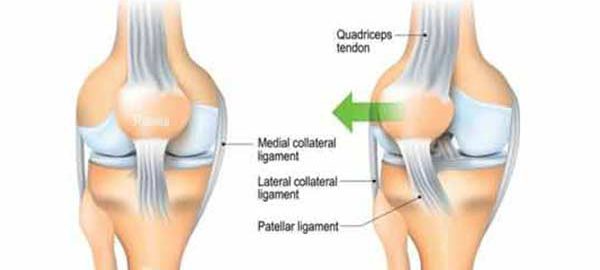
Disorders of the patella are quite common conditions, they can occur on a normal knee or previously affected by congenital diseases. From a clinical point of view, there are spontaneous or aggravated pain due to flexion of the leg, they may have variable intensity.

When the knee is inspected, the displaced patella is observed and in its place, a depression occurs. Hydrarthrosis can accompany the condition, without being important.

The treatment of luxation is orthopedic or compressive bandage can be used. In severe cases, it may be possible to reach the patellectomy. Recovery through physical therapy is mandatory.

The fractures of the patella can occur both directly and indirectly, by an exaggerated flexion of the calf.

Clinically, pain occurs, joint swelling, hemarthrosis, functional impotence. At the touch of the knee, the displaced fragments are noticed.

The treatment of fractures may vary depending on the severity. It goes from rest to bed, compressive bandage, to gypsum cast, patellectomy. The walk can be resumed after about three weeks.

The recovery of the fractured knee is made from the period of immobilization: treating inflammation, ensuring lymphatic drainage, maintaining muscle tone, recovering after the end of immobilization by passive exercises done by kinetotherapist in water, but also by active movements, reinventing the gait at the beginning with carriage and then with a cane., reconfiguration to the orthostatic position, toning of the muscles, joint mobilization and regaining the amplitude of flexion, are the steps that will have to be followed.

Prophylactic treatment is a very important one, in that the sudden movements at the knee level will be avoided by the performance athletes but also by the elderly and children who have increased sensitivity at this level.