Tag Archives: orthopaedic in Delhi

hip pain

What are hip diseases?

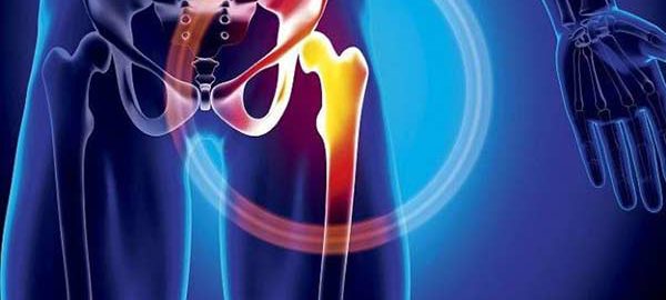
Hip pain is the main symptom that something is not right and it can be related to several diseases. 

We know that the hip is responsible for supporting the body and connecting the thigh bone (femur) to the pelvis (acetabulum). And this fitting of the head of the femur with the bone of the pelvis needs to be exact, as any type of deficiency or malformation results in the emergence of problems in the region, which include pain in the gluteus, lower back, groin, knees and legs.

It is important that the cause is always investigated so that the appropriate treatment is applied. Among the main diseases of the hip are:

  • Osteoarthritis: chronic degenerative disease that wears out cartilage, decreasing joint space, which causes limited movements, leading the person to start limping, in most cases.
  • Arthritis: Chronic inflammatory and autoimmune disease, which can affect multiple joints. It generates pain, swelling and reduced range of motion. The main cause for this pathology is genetics.
  • Epiphysiolysis: this disease is related to the growth of the femur that generates the slipping of the femoral head in the pelvis. Symptoms include groin pain, which may extend to the inner thigh to the knee, and decreased hip mobility.
  • Hip Developmental Dysplasia (DDH): known as congenital dislocation, DDH happens when the femur is not articulated with the pelvis, that is, there is a displacement between the head of the thigh bone and the acetabulum. The disease brings a noticeable difference between the limbs, in addition to causing pain in the knees and lumbar spine.
  • Bursitis: is characterized by inflammation of the bursa, a small fluid-filled sac that surrounds the joints and acts as a cushion between the bones, tendons and muscle tissues. Causes pain when getting up, walking, climbing stairs, and driving.
  • Pubalgia: tendinitis of the adductors of the hip or abdomen, with all affected muscles and tendons having their insertion in the pubic bone.
  • Femoroacetabular impingement syndrome: when there are one or more changes in the shape of the hip, which can be of the CAME, PINCER type or a combination of them (mixed type). These bone changes can cause abnormal contact between the femoral neck, acetabular rim and acetabular labrum, causing damage to the articular cartilage.

If you notice any of the symptoms listed above, it is important to seek the help of a hip specialist orthopaedic in Delhi. This is the case of Dr. Ashu Consul, who has been working in this area for over 15 years. Schedule a consultation right now!

hip arthritis

Coxarthrosis: Osteoarthritis of the Hip Joint

Coxarthrosis is osteoarthritis of the hip joint.

It is called primitive when there is no malformation responsible for osteoarthritis, and secondary in the opposite case.

The diagnosis is made based on pain and examination of the hip and is confirmed with x-rays. Treatment involves non-drug measures, medications, and eventually a resort to hip replacement surgery in Delhi.

The two coxarthrosis

The primitive coxarthrosis It occurs in a normal hip, in an individual over 60 years of age and represents 40% of the cases of coxarthrosis.

The secondary coxarthrosis it occurs in a “dysplastic” hip (with an anatomical malformation), in a younger individual, and accounts for 60% of cases of coxarthrosis.

Morphological abnormalities are, therefore, the main risk factor for coxarthrosis.

However, we must not forget the excess weight (overweight and obesity) and the trauma and microtrauma of contact sports.

Diagnosis is indicated by symptoms and examination

The symptoms are the same as those of coxarthrosis, both primitive and secondary.

Pain is the main symptom. It is a mechanical pain that is accompanied by a limp. This is usually located in the groin, but it can also be located in the buttock or reveal itself through knee pain.

Joint stiffness associated with pain is responsible for a disability that also manifests itself as discomfort when walking and lameness. The examination performed by the orthopaedic in Delhi reveals a limitation of hip movements.

Radiology confirms the diagnosis

Radiography of both hips is essential for diagnosis.

This has two objectives: search (or confirmation if it is already known) of a hip dysplasia and search for radiological signs of coxarthrosis:

  • decrease in the height of the space between the articular surfaces (impingement of the interline);
  • condensation of the part of the bone immediately under the cartilage;
  • bony outgrowths at the bone-cartilage junction (osteophytes).

Non-drug treatments take center stage

The reduction of any possible excess weight is a priority. The same happens with the implementation of an “aerobic” exercise program (30-minute brisk walk 3 times a week, cycling, swimming) and functional rehabilitation exercises in order to maintain hip mobility and strengthen the muscles that stabilize it.

The orthotics are reduced to shock-absorbing soles (running shoes with thick soles) and the technical assistance of a cane that will be carried on the opposite side to the diseased hip. The joint economy recommendations are not superfluous: avoid lifting weights, walking on rough terrain and standing for a long time.

Medications are administered in 2 ways

For the treatment of coxarthrosis, drugs are prescribed in the form of tablets or, less often, in the form of an intra-articular injection. Analgesics (paracetamol) are prescribed first.

Non-steroidal anti-inflammatory drugs have a superior analgesic effect, but they are not without undesirable effects. If there are no contraindications, they are prescribed in short cures, with effective doses, preferably during painful outbreaks.

Symptomatic slow-acting antiarthrosis drugs represent the background treatment. Its excellent tolerance authorizes its long-term prescription. Its action is delayed, but often prolonged after stopping treatment.

Intra-articular corticosteroid injections are indicated in the event of an inflammatory flare. Technique and vigilance must be rigorous.

Surgery is contemplated when disability becomes significant

The total hip replacement is the preferred prosthesis in case of coxarthrosis.

This gives good results against pain and for the autonomy of the arthritic patient.

Rheumatic-Disease

How to take care of your joints

The joint is a structure made up of bones, muscles, synovium, cartilage, and ligaments such as the knee, hip, wrist, or shoulder. They are the most important joints between the ends of the bones and allow the mobility of the skeleton.

The structures that allow this union are the joint capsule and the ligaments. Inside the capsule, the synovial membrane secretes a fluid, synovial fluid, which acts as a lubricant, facilitating the friction of the articulating bone ends. It is like oil for a car.

Another important component of the joints is the cartilage, which covers the articular bone surfaces and acts as a shock absorber in the movements of movement between them.

JOINT PAIN

Joint pain is the most common disorder affecting the musculoskeletal system. This type of pain can affect a single joint (monarticular) or several (polyarticular) and originate in the joint itself or outside of it, such as in a ligament, cartilage, tendon, or muscle.

The conditions that cause joint pain are osteoarthritis, arthritis, fibromyalgia, and gout, among others. Two of the best known are:

  • Osteoarthritis: it is the wear of the cartilage of the joint and causes the bones to rub against each other. This causes pain and loss of flexibility. 10% of the Spanish population has osteoarthritis of the knee and 6% of the hand.
  • Rheumatoid arthritis (RA):  a chronic inflammatory joint disease, which causes joint destruction, deformity, and functional limitation.

HOW TO TAKE CARE OF THE JOINTS

According to orthopaedic in Delhi, Joint care is important at all ages since, as time passes, they lose their functionality. Keep these recommendations in mind:

  1. Be active. It is advisable to practice aerobic activities such as walking, swimming, and cycling. Exercises help keep the joint active and prevent muscle atrophy. You should exercise regularly and gently to avoid injury.
  2. Correct posture. Bad posture produces an uneven distribution of weight on the body. When sitting, your back should be straight and supported on a backrest. Avoid sinking chairs, a firm seat is better; feet must be firmly on the ground.  Do not spend a long time standing or sitting, change your position from time to time. To pick up objects from the ground, do not bend your spine, but rather your knees.
  3. Control your weight.  Excess weight will put stress on your joints, especially spine, knees, hips, and feet. Obesity increases the risk of osteoarthritis.
  4. Feeding. Try to maintain a balanced diet, containing fruits and vegetables. Seasonings like turmeric are proven to reduce inflammation, oranges have antioxidant effects and protect joints, the Mediterranean diet reduces pain and stiffness in those with rheumatoid arthritis, broccoli protects against arthritis pain, and ginger is anti-inflammatory and antioxidant.
  5. Avoid overload.  Activities that generate extreme pain or exertion should be stopped or limited.  Wear wide shoes, the insoles can be used to mitigate pain in the joints of the legs. Canes and crutches can also relieve pain, by unloading diseased joints, while preventing falls.
  6. Use assistive devices.  To prevent falls, place handles on the walls, hallways, and bathrooms to facilitate holding. The taps should preferably be single lever. A trolley on wheels can allow the transport of various objects (daily groceries, dishes …) without making great efforts. In the bathroom, install non-slip floors, supports for the entry, and exit of the bathtub and toilet, plastic seats for the shower.
  7. Some compounds such as collagen and hyaluronic acid promote mobility.

calcification

Calcification: Causes, Symptoms, Prevention And Treatment of Calcium Deposits

Calcium is incredibly important for the functioning of the human body. Although 99 percent is found in teeth and bones, blood vessels, nerves, and muscles also need calcium.

They get it through blood vapor, in which that important remaining one percent must reside.

The term “calcification” refers to the accumulation of calcium in parts of the body where it should not accumulate, causing hardening of the tissues in question. This type of calcium build-up can affect many parts of the body, including:

  • The arteries ( atherosclerosis )
  • heart valves (calcification of the aortic valve)
  • other organs, such as the kidneys, bladder, and even (though rare) the liver
  • other soft tissues (muscles, breasts, fatty tissue)
  • joints and tendons.
  • the brain (cranial calcification)

What Causes Calcification?

Some calcification is a normal and expected part of the aging process. Almost all adult humans have some calcification of the pineal gland in the brain, and about half of women over 50 have some calcification within their breast tissues.

However, calcifications can also be pathological, and depending on the type of calcification, the following causes may play a role:

  • Excessive intake of vitamin D (soft tissue calcification, kidney stones)
  • a vitamin K deficiency
  • age in combination with excessive physical activity and / or being overweight (calcification as a complication of osteoarthritis)
  • genetics
  • existing heart or kidney conditions
  • taking certain medications that affect the way your body processes calcium, such as high blood pressure and cholesterol medications.
  • injury
  • of smoking

How do you know if you are experiencing calcification? Can you prevent it?

Due to the various parts of the body that can be affected by calcification, it is impossible to make general statements about the symptoms that people may experience. However, most of the time there will be no obvious symptoms, and it is very likely that you will discover calcifications after undergoing an X-ray for entirely different reasons.

The best thing anyone can do to try to prevent calcification is to live a healthy life and talk to the orthopaedic doctor in Delhi about prevention if they are at higher risk. It is also advisable that you attend preventive medical exams with your doctor, especially once you reach middle age and beyond.

Treatment for calcification

Treatment for calcification depends entirely on the type you are dealing with. Calcium deposits in the joints and tendons can be removed surgically. While people with kidney stones will likely be prescribed diuretics that stimulate calcium build-up in the bones.

Those prone to kidney stones may also be advised to reduce their calcium intake. Surgery may be necessary if you suffer from heart valve calcification, while those with cerebral calcification may be prescribed bisphosphonates, a class of medication that is also used to treat osteoporosis.

In conclusion

While calcification of any kind is unlikely to produce symptoms that easily lead to self-diagnosis, anyone concerned about their health should see a orthopaedic in Delhi. The detailed description of your symptoms will allow your doctor to decide what kinds of tests should be done, leading to a diagnosis.

carpal-tunnel-syndrome

Carpal Tunnel Operation

Carpal Tunnel Operation duration

The duration of the Carpal Tunnel Operation is approximately 60 minutes. Generally, the patient can go home the same day of the surgery, although it will depend on the indications of the orthopaedic doctor in Delhi.

What is the Carpal Tunnel Operation?

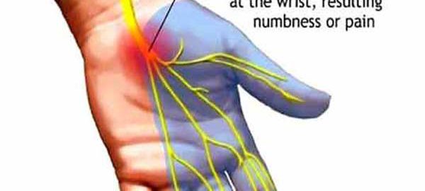
The carpal tunnel syndrome is a condition in which the median nerve is subjected to excessive pressure. The median nerve is located in the wrist and allows the hand to have sensitivity and movement in certain parts. When a person has carpal tunnel syndrome, they often feel numbness, tingling, weakness, or muscle damage to the hand and fingers.

This syndrome can be caused by using tools that vibrate and it is even believed that other activities such as typing, using the mouse, playing an instrument … can cause this inflammation and narrow the carpal tunnel, causing pain.

Other causes can be alcoholism, fractures of the bones, arthritis in the wrist, cyst or tumor in the wrist, infections, obesity, fluids that accumulate during pregnancy and menopause, or rheumatoid arthritis.

It is more common in women than in men and occurs especially in people between 30 and 60 years old.

Surgery Information

What is Carpal Tunnel Release Surgery?

It is a minimally invasive surgical procedure in which the ligament that exerts pressure on the nerve is cut. Local anaesthesia is used and the patient leaves the hospital the same day. At home, the patient is advised to make hand and wrist movements to avoid stiffness and swelling.

The surgical intervention has a very short duration, about 15 minutes.

The use of a special wristband may be indicated during the first three weeks. Of course, overexertion and forced wrist postures should be avoided.

Normally it is a very effective surgery, but it will depend on each patient. Full recovery can take about two months.

After the operation you may feel discomfort or swelling, for this, the orthopaedic in Delhi will prescribe the indicated medication.

Full recovery usually occurs after these 4 or 6 months. This intervention is usually very effective. The innovative techniques used make possible a faster recovery and fewer complications in surgery.

Do not hesitate to go to a consultation with the orthopaedic surgeon in Delhi if you think you may have this problem.

What symptoms does carpal tunnel syndrome have?

Some of the most common symptoms are:

  • The clumsiness of the hand when grasping objects.
  • Numbness and tingling in the thumb and the next two or three fingers. It can also be felt in the palm of the hand.
  • Pain from hand to elbow.
  • Pain in the hand or wrist.
  • Problems with coordination in fine movements.
  • Atrophy of the muscle below the thumb.
  • Difficulty loading bags.
  • Weakness in one or both hands.

What is the initial treatment?

When this syndrome appears, your orthopaedic doctor in Dwarka will advise you to wear a splint at night, avoid sleeping on your wrists and apply cold or heat to the affected area. Additionally, nonsteroidal anti-inflammatory drugs (NSAIDs) or corticosteroid injections may be prescribed to relieve symptoms.

When none of these treatments works, carpal tunnel release surgery can be used. In the long term, surgical treatment is the only solution for carpal tunnel syndrome.

elbow-fracture

What to do with an Elbow Fracture

They are frequent in children and can become complex.

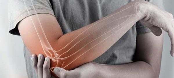
The fractures of the elbow include a wide spectrum of injuries whose treatment ranges from a brief period of immobilization and immediate rehabilitation to complex surgery and long periods of rehabilitation.

Dr. Ashu Consul an elbow and shoulder specialist traumatologist, orthopaedic surgeon in Delhi, indicates that when faced with a traumatic elbow injury, the first thing to do is apply ice, immobilize with a sling and consult an emergency or trauma service to Clinically and imaging the lesion.

“In those minor fractures, for example, fractures of the radius dome without displacement, a short immobilization period of 7-10 days is sufficient, and then early mobilization begins, sometimes assisted with kinesiology depending on the injury, ”orthopaedic in Delhi explains.

Above all, in children, elbow fractures are frequent, especially those that involve the distal humerus says the orthopaedic doctor in Delhi. “Its management ranges from plaster for 3 weeks in those nondisplaced fractures, and surgery with reduction (alignment of the fracture) and needle fixation in those displaced fractures,” he indicates.

There are severe elbow injuries that occur in higher energy trauma and these involve severe bone or soft tissue (ligament) injury. “These types of injuries may require a complex plate and screw fixation surgeries and long periods of rehabilitation,” says Dr. Consul.

The elbow is a joint in which stiffness is frequently presented as a complication, which is why its management must be carried out by specialists, both trauma and kinesiologists, he emphasizes.

Full and definitive recovery from a complex elbow fracture can take 6 to 8 months.

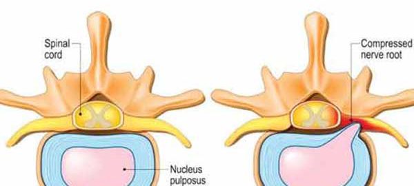
Disc-Herniation

Everything you need to know about Disc Herniation

Disc herniation means the movement of discs in the spine. As a result of this condition, intense back pain occurs in that area.

Depending on the area of ​​the back where they occur, the most common types of herniated disc are lumbar and cervical.

Lumbar disc herniation occurs when the movement of the intervertebral disc has occurred at the lumbar level. The most affected are the last two lumbar discs, L4 and L5, due to the fact that they support most of the body weight. The most affected nerves are the sciatic and femoral.

Cervical disc herniation occurs when the displacement of the intervertebral disc has occurred in the upper part of the spine.

Causes

Disc herniation is most often the result of gradual wear and tear related to aging, called pulpal nucleus degeneration. As a person gets older, the discs lose some of their fluid content. This makes them less flexible and more prone to cracking or breaking, even in a small movement or twist.

Predisposition to sedentary lifestyle, prolonged sitting, increased exertion after a longer period of rest, lifting weights incorrectly, but also spinal cord injuries are among the causes of disc herniations, explains the orthopaedic in Delhi.

Risk factors

Weight. Excessive weight causes extra stress on the lower back discs.

Occupation. People with high-intensity jobs are at higher risk of developing spinal problems. Lifting, pulling, or pushing weights, repeated movements, or lateral bending and twisting may increase the risk of a herniated disc.

Genetics. Some people inherit a predisposition to the development of a herniated disc.

Symptoms

The symptoms of a herniated disc differ depending on the area in which it occurs.

In the case of lumbar disc herniation, there is pain in the thighs and buttocks, which can radiate to the leg below the knee. Pain may be accompanied by tingling, numbness and tenderness in the limbs and affected areas.

In the case of cervical hernia, the pain manifests itself mainly in the shoulders and arms. There is pain in the neck region, with the impossibility of turning the head or making sudden movements.

The pain may be accompanied by tingling, numbness and a general state of weakness. In both cases, the pain is continuous and not throbbing.

Treatment

Disc herniation treatment should not be delayed because the pain subsided at some point. Lack of treatment can worsen the situation, causing chronic pain or permanent injuries that affect mobility, says the orthopaedic surgeon in Delhi.

Physiotherapy is the most common methods of treating a herniated disc. In severe cases, when the pain persists even after 5-6 weeks of alternative treatment, surgery is resorted to.

The operation consists of removing the fragments of the disc or even the entire herniated disc that compresses the spinal nerves.

hip-joint-pain

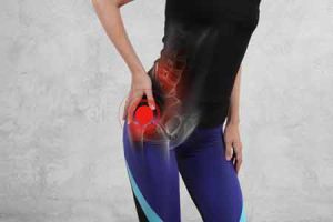
The Main Causes That Influence Hip Pain

When you tell your orthopaedic in Delhi that your hip is hurting, the first thing the doctor needs to do is confirm that the balance is the problem.

Women can say they have hip pain, but they can also say that they have pain in the upper thigh or upper buttock.

These hip pains may be faced with minor back pain. Hip pain can be felt very often in the area of ​​the abdomen or the outer part of the hip over which its joint is located.

Causes of hip pain

If a female patient complains of hip pain, the orthopaedic in Dwarka firstly considers the patient’s age and level of activity. Among the most common causes of hip pain in women, we mention:

  1. Arthritis. The most common causes of chronic hip pain in women are arthritis, especially osteoarthritis, which affects many people as they get older. Arthritis pain is most often felt in the front of the thigh or in the abdomen area, due to stiffness or swelling.
  2. Hip fractures. Hip fractures are common in older women, especially those with osteoporosis (decreased bone density). Symptoms of a hip fracture include severe pain.
  3. Tendonitis and bursitis. Many tendons around the hip connect the muscles with each other. These tendons can easily become inflamed if you overload them or participate in intense physical activity. One of the most common causes of tendonitis in the hip joint, especially for athletes, is the iliotibial tract syndrome. Another common cause of hip pain in women is exchange. There are some liquid bags in the bony side of the hip, called bursa pillows. Like tendons, these bags may become inflamed from irritation or over-stress, causing pain every time the hip joint moves.
  4. Hernia. In the abdomen area, the groin and femoral hernias – sometimes referred to as sports hernias – can cause anterior (frontal) pain in the woman’s hip. Pregnant women may be sensitive to groin hernias due to the pressure added to the wall of their abdomen.
  5. Gynecological problems. Hip pain in women can also have gynecological causes. It is important not to just realize that the pain can be caused by arthritis, bursitis or tendonitis. Depending on the age or other health problems, hip pain may also come from another system. Endometriosis can cause pelvic sensitivity, so some women describe this problem as hip pain.

Back pain or spinal pain can also be mentioned and felt around the buttocks or hip. Sciatica can cause pain in the back of the hip – sciatica pain can start in the lower back and reach the buttocks and even the legs.

Arthritis is a common cause of hip pain and mobility change. More than a quarter of older adults develop coxarthrosis pain, which threatens mobility – slower walking, difficult stairs. The term “arthritis” is covered under a number of different conditions, including osteoarthritis and inflammatory diseases, such as rheumatoid arthritis and psoriatic arthritis. If you find out what kind of arthritis you have, you must be able to successfully reduce hip pain. And finding out the type of arthritis is a solution that reduces your hip pain. The most common type of arthritis is osteoarthritis. Osteoarthritis can also result from a common injury, sometimes called traumatic arthritis.

Factors that increase the risk of osteoarthritis include:
1. Aging;
2. Obesity;
3. Deterioration or various injuries in the joint;
4. Structural problems with the joint;
5. Rheumatoid arthritis.

Symptoms of osteoarthritis develop slowly, starting with stiffness or pain in one or both bones of the hip and eventually becoming painful enough to prevent various normal activities (walking or climbing stairs).

Rheumatoid arthritis can also be a problem that causes pain. Inflammation may occur due to an abnormality of the immune system. Rheumatoid arthritis is a chronic disease that can affect the entire body, not just the hips. It starts with a swelling of the mucosa of the joints, called the synovial mucosa and progresses to the deterioration of bones and cartilage. The cause of rheumatoid arthritis is not fully understood, although an abnormal response of the immune system to the body contributes to the disease.

Symptoms of rheumatoid arthritis also include:
a. Pains that increase slightly in intensity in the symmetrical joints;
b. Swelling of the affected joints;
c. Fatigue;
d. morning rigidity;
e. Pain after a period of stay;
f. Weakness;
g. Muscle pain ;
h. Anemia.

Early diagnosis of rheumatoid arthritis is critical to maintaining the quality of life. Medications can slow the progression of this disease. A less common type of arthritis that causes joint pain is psoriatic arthritis, a complication of psoriasis, inflammatory skin disease.

Strategies to ease discomfort:

  • Lose weight if you are overweight in order to ease the weight of the joints;
  • Try analgesics, but be careful about recommended doses or warnings;
  • Ask your doctor for more information about common supplements such as glucosamine and chondroitin.
  • Work with a physical therapist to do stretching and flexibility exercises;
  • Use ice and heat to relieve painful areas.

If your mobility is impaired, ask your orthopaedic in West Delhi if you would like the assistance of a support device to help you with walking or other daily activities. Discuss in detail the options for prescription drugs, injections, or surgeries with the orthopaedic surgeon in Delhi so that a treatment plan is tailored to your needs.

Managing hip pain

Things you can do to better manage your hip pain:

  1. Exercises – do exercises in the morning to put your muscles to work, to be active. Press on your ankles and lift your buttocks off the floor as you tighten your abdominal muscles. Keep your knees aligned with your ankles and follow a straight line from your knees to your shoulders, without bending your back. Stay in that position for three to five seconds. Start with a set of ten exercises.
  2. Use ice – against arthritis or bursitis ice can be used, thus reducing inflammation and helping against hip pain. Use an ice pack, wrap it in a towel and place it where you feel the pain.
  3. Use heat for arthritis – warming an arthritic balance with a hot shower or bath can relieve pain. Do not use heat, if hip pain is caused by the stock market.
  4. Strengthening the thighs – this is another muscle group that contributes to supporting the hips. Lie on your back, place a ball between your knees and tighten. And a hard pillow or Pilates ring will help. Start with a set of ten repetitions and proceed with approximately up to three sets. Then strengthen your thighs on the outside.
  5. Work out in the water – swimming or aerobics in water are wonderful exercises for the hip joints. Exercises in water allow you to strengthen your muscles without putting pressure on your joints.
  6. Avoid activities that have a high impact – jumps can cause hip pain to worsen. Walking is a better choice.
  7. Weight loss – if you have osteoarthritis in the hip that results from cartilage wear, losing even a few pounds can help against joint and hip pain.
  8. Listen to your body – if you have arthritis or tummy tuck, you may have noticed that exercise can help relieve pain. If the balance starts to hurt during certain exercises and persists longer, this is a sign that you need rest and that you should rest. It is normal to feel pain one day after exercise, but the pain should not persist or become worse. Also, if you experience sharp pain, stop your activity and talk to a doctor or a physiotherapist in Delhi.

If you undergo major hip replacement surgery in Delhi, it is very important to prepare yourself in advance to increase your chances of an optimistic outcome and reduce hip pain. The most important thing you can do before surgery is to make sure you are on the same wavelength as the orthopedic surgeon. You need a doctor to talk to and gain confidence in, so you get a sense of well-being. Before replacing your balance, you will be evaluated by your orthopaedic surgeon in Dwarka to make sure you are healthy enough for the operation.

This evaluation could include an electrocardiogram, chest x-ray, and various blood tests. Your doctor will use this information to determine if you have an increased risk of complications. Note that certain herbal medicines, including supplements, may increase the risk of bleeding and other serious complications during surgery. So make sure that both your orthopaedic surgeon in West Delhi and you are aware of all the medicines, herbal supplements you are administering.

Balance replacement is a major operation and there is the possibility of needing a blood transfusion during or after the operation. However, you can start donating blood about four weeks before surgery. Inform yourself by asking your orthopedic in Delhi what you should do about how much blood can be stored safely before surgery.

To produce more red blood cells (this requires an injection once a week for about a month before surgery), you can also take other medicines (epoetin alfa). In this way, patients have an extra reserve in terms of blood supply when they undergo surgery.

Men with prostate problems should be checked by a urologist prior to surgery. After you schedule your hip replacement in Delhi, call your dentist. If you have problems with your teeth, you should go for a checkup. Infections from an abscess or other dental infection can spread to the hip, causing serious complications.

Cuboid

Leg Injuries – Cuboid Syndrome

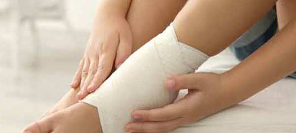
Cuboid syndrome is the result of injuries to the joints and ligaments surrounding the cuboid bone. This is one of the seven tarsal bones of the foot (talus or astragalus, calcaneus, navicular, cuboid and 3 cuneiform bones).

From a medical point of view, the condition is called a cuboid subluxation and occurs when the cuboid bone moves down and out, without being aligned with the calcaneus bone.

The cuboid syndrome causes pain in the external side of the foot, which can be confused with other conditions.

Clinical picture

Cuboid syndrome encompasses a variety of common symptoms and other conditions, sometimes making it difficult to diagnose. The most common symptoms include:

  • Pain on the outside of the foot, which affects including fingers 4 and 5;
  • Pain that aggravates the standing state, due to body weight ;
  • Painful pain, dry or sharp and acute;
  • Weakness and difficulty in walking and jumping;
  • Redness, inflammation, and sensitivity in the injured area;
  • Walking painkillers, to reduce pain;
  • Reducing the range of movements of the foot and ankle.

Studies have reported that, in general, the cuboid syndrome is not uncommon in the population, but occurs most commonly in athletes and dancers. When the cuboid syndrome is correctly identified and treated, most people experience a complete recovery.

Causes

Cuboid syndrome can have different causes, but the most common are:

  • Overload – this condition, along with injury, is most commonly seen in athletes and dancers. These groups of athletes tend to have intense physical activity, regardless of the presence of pain. Overload injuries tend to develop after long periods of intense activity, such as running;
  • Ankle dislocation – ankle dislocation can also lead to injury to the Tarsus bones of the foot. A specialist study reported that about 40% of people with ankle dislocations or sprains can develop cuboid syndrome;
  • Foot walking in pronation – people who develop this type of walking have a higher risk of having the cuboid syndrome because the muscles of the leg can pull the tarsal bones into abnormal positions;
  • Other causes – climbing, uneven surfaces and sports involving fast and lateral movements, such as tennis, can cause the cuboid syndrome. Wearing shoes of poor quality or inadequate sizes may be another reason.

Also, if you do not take into account the indications of orthopaedic in Delhi regarding rest and recovery from a leg injury, various complications, including cuboid syndrome, may occur.

Diagnostics
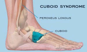

The foot represents a complex, flexible and resistant segment of the body. It contains about 100 muscles, ligaments and tendons, 28 bones and 30 joints.

The complex structure of the foot and the non-specific nature of the pain within the cuboid syndrome make this injury difficult to diagnose.

Sometimes, medical imaging techniques such as X-ray or magnetic resonance imaging (MRI) do not identify signs of this condition, even if it is present. The cuboid syndrome may also mimic the symptoms of other foot problems, such as fractures or sprained heels.

In order to establish a correct diagnosis and to find the most effective treatment, the physician must perform a thorough physical examination and examine a person’s medical history.

Treatment options

Treating cuboid syndrome always involves rest and elimination of activities that involve putting pressure on the injured leg. Applying ice and an elastic phase, but also raising the foot above the level of the heart on a pillow are other useful methods.

If the pain persists or worsens, the orthopaedic in Dwarka should be consulted urgently. Certain maneuvers performed by the therapists in the field can help to improve or eliminate the symptoms.

These maneuvers are meant to force the bone to return to its original position. However, it should be noted that such maneuvers are not recommended if there are other associated conditions, such as arthritis, bone disease, fractures, circulatory or nerve disorders.

Additional treatments may include the use of orthotics to support and properly align the foot, but also to administer anti-inflammatory drugs to reduce pain and inflammation.

In some cases, leg massage can help to reposition the bone. Surgery is used in rare cases when other treatment options have not worked.

Recovery

The length of time required for recovery from cuboid syndrome depends on several factors, such as:

  • Duration of the treatment until the start of treatment;
  • Cause of cuboid syndrome ;
  • If the initial injury was mild or severe;
  • If the instructions of orthopaedic in West Delhi were taken into account;
  • If the condition has been correctly diagnosed and treated.

Physiotherapy in Dwarka can play an important role in the complete recovery of a person with the cuboid syndrome. Also, this therapy can prevent further complications.

Physical therapy includes:

  • Reconsolidation of the strength and function of the foot;
  • Maintenance of the muscles;
  • Exercises to improve balance.

In some cases, to avoid pressure on the injured leg, the orthopaedic surgeon in Delhi may indicate the use of squats or walking sticks.

Risk factors

The common risk factors for the cuboid syndrome are:

  • Overweight or obesity;
  • Wearing shoes of inappropriate sizes;
  • Wearing pairs of shoes of inferior quality;
  • Practicing physical exercises without adequate heating;
  • Practicing physical activities without taking into account pain and without having sufficient rest periods;
  • Performing activities, such as running or walking on uneven or rough surfaces;
  • Fractures of neighboring bones of the cuboid;
  • Practicing sports such as athletics or ballet;
  • Practicing physical activities that involve repetitive movements and increased impact on the foot;
  • Association of other conditions such as arthritis or osteoporosis.

Perspective

Cuboid syndrome is not a serious condition and can be easily treated by the orthopaedic surgeon in Dwarka, sometimes involving physical therapy. The perspective is usually very good for people with this condition. Following treatment, most people can return to normal activities, with little risk of recurrence.

The orthopedic in Delhi should be notified if persistent pain occurs in the foot before doing any maneuver that could aggravate the situation.

Often there may be other health conditions that mimic cuboid syndrome. Rapid identification and early treatment of health status offer the best chances of recovery.

meniscus lesions of knee joint

Meniscus Lesions of The Knee Joint

The two meniscus can be easily injured by a rotation of the knee during a sustained effort. Partial or total meniscus rupture occurs at an abrupt torsion or thigh rotation, while the foot stays in place (for example, at a sudden turn to hit the tennis ball).

If the rupture is minimal, the meniscus remains connected in the anterior and posterior part of the knee; if the rupture is large, the meniscus may hang from a cartilage filament. The severity of a rupture depends on the location and extent.

Symptoms

In general, pain in the meniscus rupture occurs, especially when the leg is straightened (at the extension of the leg). The pain may be moderate and the patient may continue the activity.

Severe pain occurs when a fragment of the meniscus remains between the femur and tibia. Swelling may occur immediately after injury if the blood vessels are damaged, or a few hours after injury if the joint space is filled with fluid produced in response to inflammation. If the synovial envelope is damaged, it becomes inflamed and produces fluid to protect itself, installing the synovial envelope syndrome.

These changes cause swelling of the knee. Sometimes an untreated injury can become painful after several months or even years later, especially if the knee has been injured a second time. After an injury, the knee may become blocked or weakened. Symptoms of a meniscus lesion may resolve spontaneously, but frequently the symptoms persist and require treatment.

Diagnostic

In addition to the patient’s anamnesis, which describes the onset of pain and swelling, the orthopaedic in Dwarka may perform an x-ray of the affected knee. The examination may include a test in which the doctor flexes the leg, then rotates internally and externally while extending the leg. The pain from this maneuver suggests the meniscus rupture. An imaging magnetic resonance test may be recommended to confirm the diagnosis. Occasionally, the orthopaedic surgeon in Dwarka may use arthroscopy for diagnosis and treatment.

Treatment

If the injury is minimal and the pain and other symptoms disappear, the orthopaedic in Delhi will recommend a program of exercises to strengthen the muscles. Exercises for meniscus disorders are initially performed under the supervision of an orthopaedic in West Delhi or therapist.

The physiotherapist in Dwarka will ensure that the patient performs the exercises correctly and without the risk of a new injury. The following exercises are intended to strengthen the thigh muscles and increase the elasticity:

  • warming the joint by walking on a medical bicycle, then stretching and lifting the leg
  • extension of the foot from the sitting position (a small weight can be placed on the ankle for this exercise)
  • in ventral decubitus, there is performed leg lifting movements
  • exercises in the swimming pool, including the rapid walking with water at the chest level, raising each leg to 90 with the back glued to a wall of the pool.

If the rupture is enlarged, the doctor may perform arthroscopy in Delhi or surgery to assess the severity and repair the lesion. The orthopaedic surgeon in Delhi can suture the meniscus in relatively young patients if the lesion is in a well-vascularized area and the ligaments are intact. Most young athletes will be able to perform vigorous sports activities after meniscus repair.

In elderly patients or if the rupture is in a poorly vascularized area, the doctor will resect a small portion of the meniscus to smooth the surface. In some cases, the orthopaedic surgeon in West Delhi will resect the entire meniscus.

However, degenerative rheumatism, such as osteoarthritis, presents a higher risk of development if the meniscus is completely removed. Researchers are investigating a procedure by which the meniscus is replaced with a meniscus from a corpse (allograft). A meniscus graft is fragile and can shrink and break easily. The researchers tried to replace the meniscus with an artificial one, but the procedure was even less successful than the allograft.

The rehabilitation after the surgical repair of the meniscus lasts for several weeks, and the postoperative activity is more restricted than in the case of the total resection of the meniscus.

However, exercises speed up healing. Regardless of the type of surgical operation, rehabilitation includes gait, flexion and extension exercises of the knee, and exercises for strengthening the thigh muscles.

The best results of treatment for a meniscus lesion are obtained in patients without modifications of the articular cartilage and without lesions of the anterior cruciate ligament intact.